Magnetic Insight is a developer and manufacturer of preclinical Magnetic Particle Imaging (MPI) systems for use with in vivo models.
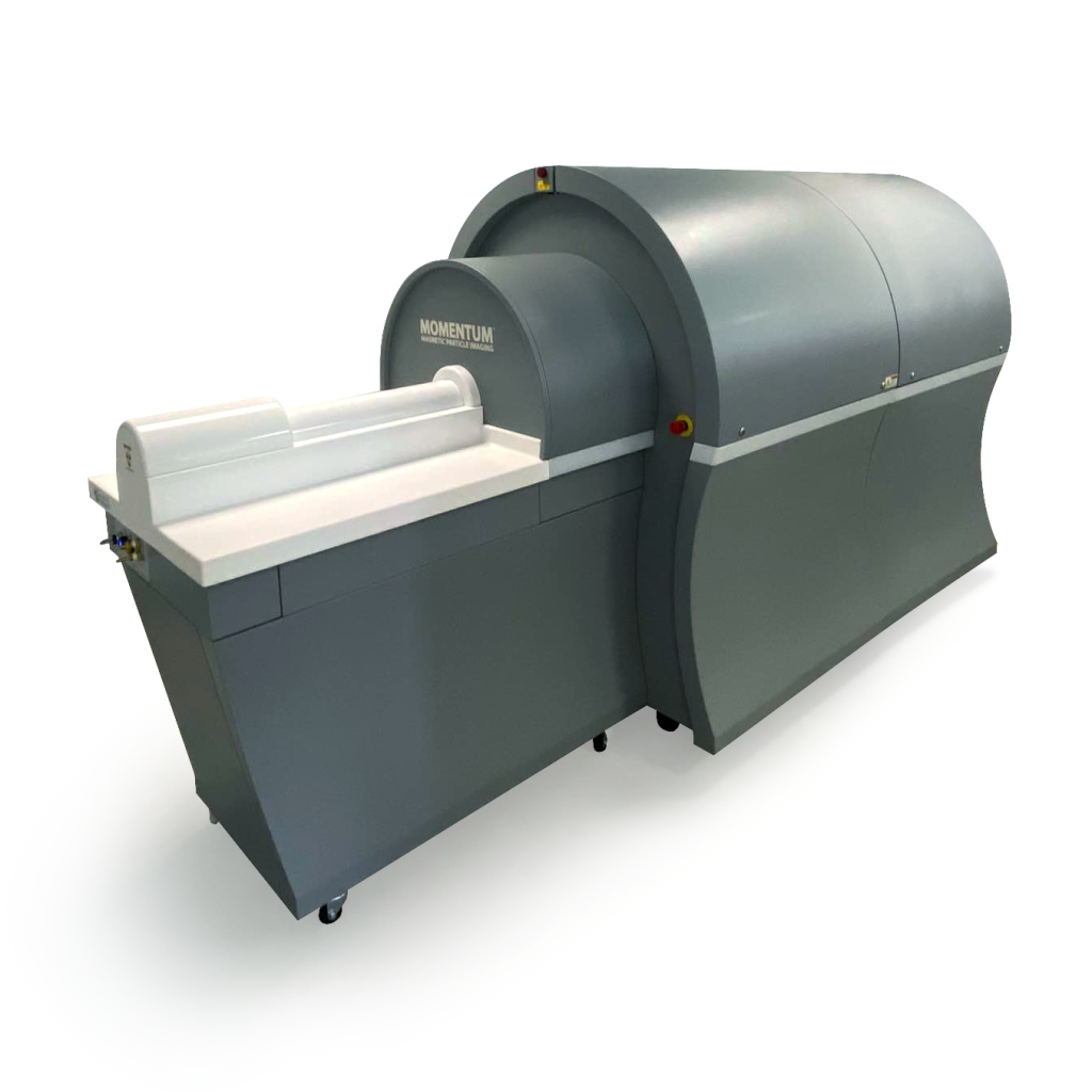
 For more information, explore our product offerings:
Momentum CT
Market-leading MPI imager, now with integrated X-ray/CT
→ View Hardware
VivoTrax and VivoTrax Plus
MPI-optimized tracers for multiple applications, including cell tracking
→ View Reagents
Relax
MPI relaxometry software option for the Momentum CT MPI System
→ View Software
MagImage
Image processing and analysis software for MPI included with our systems
→View Software
Consumables
Purchase reagents or replacement beds
→ Shop
Support
Submit a request for help with your imager or reagents.
→ Contact Support

Emerging Applications
MPI enables preclinical research applications that give a window into the behavior and treatment of illnesses, seeing processes currently invisible to conventional imaging techniques. As the MPI field expands, the list of potential new applications of MPI continues to grow. Moreover, Magnetic Insight’s active development of clinical MPI means that preclinical applications are eminently translatable in the future.
Cellular Imaging
Cellular imaging enables detecting, tracking, and quantifying cells in vivo. Over time, it allows for studying cellular events in disease processes and may be used as a biomarker to help make decisions about treatments and to monitor treatment response. The need for cellular imaging is rapidly expanding within modern medicine, with numerous applications in immunotherapy monitoring and regenerative medicine.
By tagging therapeutic cells with iron oxide tracers before administration or by tagging cells in the body through specialized tracers, MPI gives a new window into understanding cell behavior. A combination of high sensitivity to the tracer and lack of tissue attenuation means as few as 250 labeled cells can be detected.1-3 This enables clinicians to:
- Visualize cell location
- Validate cell administration
- Assess treatment status

MPI is highly quantitative, allowing for accurate measurement of the number of cells present in a region.

MPI signal from a titration of pelleted macrophage cells from 1K – 1M cells.
Pushing the Boundaries of Inflammation Monitoring
Inflammation is a part of the body’s response to harmful stimuli, such as damaged cells, irritants, and pathogens. Detecting and monitoring inflammation in a patient’s body is essential for diagnosing disease, but obtaining quantitative data is difficult using current imaging techniques such as MRI, CT, and nuclear medicine.
Magnetic nanoparticles can be loaded into various immune cells (such as monocytes and other leukocytes), then tracked as they migrate and accumulate in the region of inflammation. The nanoparticles can also be directly injected, where they are captured by phagocytotic immune cells. The inflammatory burden can be measured when these cells home to sites of inflammation.
These regions can be monitored for days to weeks using MPI, providing a quantitative measurement of inflammatory burden. This data will allow clinicians to include the inflammatory response in disease staging, helping diagnose conditions from strokes and brain injuries to autoimmune diseases like MS and arthritis.

25K RAW264.7 cells (macrophages) administered intracranially and imaged with MPI.

Iron loaded cells infiltrating a tumor are visualized ex vivo with the Perls Prussian blue (PPB) stain.
Regenerative Medicine
The restorative effects of regenerative medicine rely on the arrival and survival of therapeutic stem cells at a disease site. While experimental results have shown that cell therapy can succeed, the field has struggled to produce consistent clinical outcomes. With MPI, we can determine if intermediate steps in the treatment process succeed before seeing a change in outcome. For example, imaging nanoparticle-tagged stem cells with MPI can assess their biodistribution after cell administration, ensuring the required number of stem cells reach and are retained in the target tissue4-8. This enables:
- Measuring changes in stem cell grafts over time,
- Ensuring delivery of cells to the correct tissues,
- Improving understanding of transplant rejection.
New Possibilities in Perfusion Imaging
There is a need for more effective diagnostic perfusion imaging approaches for diseases like stroke and heart disease. Current vascular perfusion imaging techniques suffer from radiation exposure, safety, speed, sensitivity, and specificity limitations that hold back their diagnostic quality. MPI can add to the neuro and cardio interventional suite by providing a safe, rapid 3D perfusion without ionizing radiation or toxic tracers. MPI produces no signal from overlying tissues, which enables positive contrast images and real-time perfusion with unprecedented contrast-to-noise and signal-to-noise. The high contrast tracer imaging may also enable simultaneous blood vessel lumen diameter assessment and quantitative perfusion imaging.
Rat head blood pool image showing arteries, veins, and tissue perfusion
New Applications
Our customers continue to develop new applications in nanoparticle technology, hyperthermia, functional imaging, and many more.
References:
- Zhou et al. Magnetic particle imaging for radiation-free, sensitive and high-contrast vascular imaging and cell tracking. Current opinion in chemical biology2018; 45: 131–138.
- Song et al.Janus Iron Oxides @ Semiconducting Polymer Nanoparticle Tracer for Cell Tracking by Magnetic Particle Imaging. Nano Lett. 2018; 182-189.
- Zheng et al. Magnetic particle imaging tracks the long-term fate of in vivo neural cell implants with high image contrast. Sci Rep. 2015; 5: 14055.
- Gaudet et al. Imaging Cancer Immunology: Tracking Immune Cells in vivo with Magnetic Particle Imaging. J Immunol. 2019;202(1): 130-7.
- Zheng et al. Quantitative magnetic particle imaging monitors the transplantation, biodistribution, and clearance of stem cells in vivo. 2016; 6(3): 291-301.
- Nejadnik et al. Ferumoxytol can be used for quantitative magnetic particle imaging of transplanted stem cells. Mol Imaging Biol. 2019;21(3): 465-472.
- Lemaster et al. A trimodal contrast agent for stem cell tracking combining ultrasound, photoacoustics, and magnetic particle imaging (Conference Presentation). Reporters, Markers, Dyes, Nanoparticles, and Molecular Probes for Biomedical Applications XI. Vol. 10893. International Society for Optics and Photonics. 2019.
- Wang et al. Magnetic particle imaging of islet transplantation in the liver and under the kidney capsule in mouse models. Quant Imaging Med Surg. 2018;8(2): 114-122.